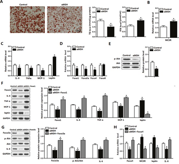
Figure 3. FoxOs abolish the suppression of αMSH on inflammation in mice adipocytes.

(A) Oil Red O staining for differentiated primary adipocytes isolated from epididymal white adipose tissue after administration of αMSH for 1 h (left). Relative concentration of TG in adipocytes (middle) and FFA in cell culture medium (right) were detected (n=3). With αMSH treatment, mRNA levels of MC5R (B), IL-6, TNFα, MCP-1 and Leptin (C), Foxo1, Foxo3a, Foxo4 and Foxo6 (D), protein levels of p-AKT and total Akt (E) were detected in adipocytes (n=3). (F) Adipocytes treated with pAd-Foxo1 and αMSH, protein levels of IL-6, TNFα, MCP-1 and Leptin were measured (n=3). (G) Expression levels for Foxo3a, p-Akt, Akt and IL-6 protein after cells treated with pAd-Foxo3a and αMSH (n=3). (H) Normalized mRNA levels of Foxo4, MC5R, Leptin and IL-6 with pAd-Foxo4 and αMSH treatments (n=3). Values are means ± SD. vs. control group, * P < 0.05.
