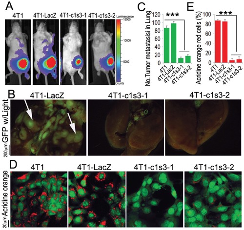
Figure 2. ATP6v1c1 knockdown in 4T1 tumor cells inhibits 4T1 tumor cells metastasis by impairing V-ATPase function.

A. Representative luciferase imaging of mice inoculated with 4T1 cells with without ATP6v1c1 knocked down by lentiviral shRNA (4T1-c1s3-1 or -2) compared to uninfected (4T1) and control infected cells (4T1-LacZ) at the primary site at day 6 after orthotopic xenograft (n=5, 4-6 weeks of age, female). B. Representative epifluorescent imaging of spontaneous tumor metastasis development in the lungs of mice with or without ATP6v1c1 knockdown in eGFP labeled cancer xenograft cell lines per observation of the lungs 22 days post mammary fat pad xenograft (n=5, 4-6 weeks of age, female). C. Quantification of metastases from B. Results are mean ± s.e.m. *P<0.05, **P<0.01, ***P<0.001. D. Effects of ATP6v1c1 knockdown on acidification at the cell membrane as indicated by acridine orange (AO) staining of different 4T1 cells treated as indicated. The cells shown were representative of the data (n=3) (primary magnification ×100). E. Quantification of acridine orange low pH staining from D. Results are mean ± s.e.m. *P<0.05, **P<0.01, ***P<0.001.
