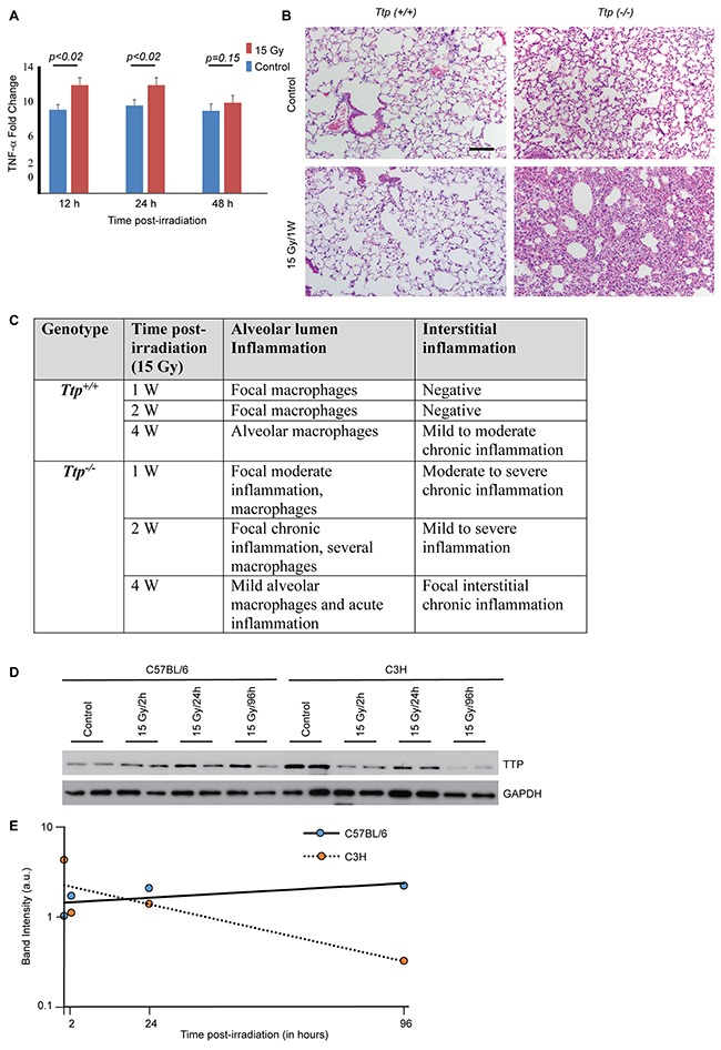
Figure 1. Ttp knockout mice are susceptible to radiation-induced lung inflammation.

(A) Fold change in TNF-α in BAL either from sham-irradiated (control) or 15 Gy single exposure of Ttp−/− mice compared to Ttp+/+ after indicated times post-irradiation (n=6 mice for each time point for each genotype category). (B) The thoraxes of Ttp+/+ or Ttp−/− mice were either sham-irradiated (- RT) or exposed to 15 Gy. Shown is representative H&E staining of paraffin-embedded lung sections. Scale bar, 200 μm. (C) Summary of histopathological observation in Ttp+/+ and Ttp−/− mice upon 15 Gy whole thorax irradiation (n=5 mice for each time point for each genotype category). (D) C57BL/6 and C3H mice strains were either sham-irradiated or 15 Gy single exposure of upper thorax. Lung specimens were collected after 2, 24 and 96 hours post-irradiation and cryo-preserved. Following completion of the study, cryosections were subjected to protein isolation as described in the materials and methods and subjected to immunoblotting for total Ttp protein. GAPDH was used as loading control. (E) Average band intensity (arbitrary units) was calculated from two representative samples from each group (as shown in panel D) using Image J software and plotted against time post-irradiation.
