
Figure 1.
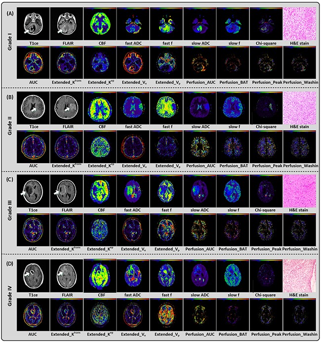
Conventional/multi-parametric MRI maps and H&E stain results of 4 individuals diagnosed as grade I (A), II (B), III (C), and IV (D) gliomas, respectively. For each individual, 1 parametric map derived from 3D ASL (i.e. CBF), 5 parametric maps derived from multi b-value DWI (i.e. fast ADC, fast f, slow ADC, slow f and Chi-square maps), part of parametric maps derived from DCE (9 out of 24, i.e. AUCAIF, Extended_Krans, Extended_Kep, Extended_Ve, Extended_Vp, Perfusion_AUCFP Perfusion_BAT, Perfusion_Peak, and Perfusion_Washin) and H&E stain (i.e. haematoxylin and eosin) result were shown.