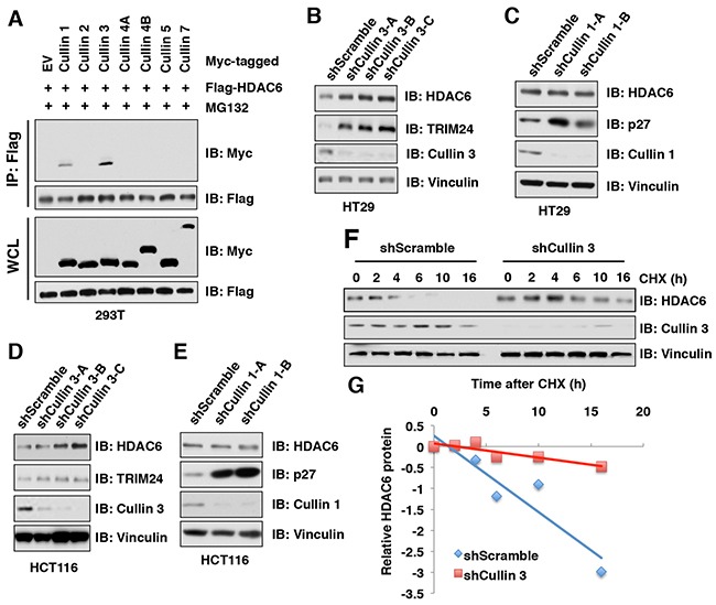
Figure 2. HDAC6 protein stability is negatively regulated by the Cullin 3 family E3 ligase.

(A) IB analysis of WCLs and immunoprecipitates (IP) derived from 293T cells transfected with Flag-HDAC6 and Myc-Cullins constructs as indicated and treated with 10 μM MG 132 before harvesting. (B-C) IB analysis of WCLs derived from HT29 cells infected with the indicated lentiviral shCullin 3 (B) or shCullin 1 (C), respectively. (D-E) IB analysis of WCLs derived from HCT116 cells infected with the indicated lentiviral shRNAs against Cullin 3 (D) or Cullin 1 (E), respectively. (F-G) IB analysis of WCLs derived from HT29 cells stably infected with the indicated lentiviral shRNAs and treated with 100 μg/ml cycloheximide (CHX) for indicated times (F). Quantification of the band intensities of (F) using the ImageJ software (G). HDAC6 immunoblot bands were normalized to Vinculin, then normalized to the t = 0 time point.
