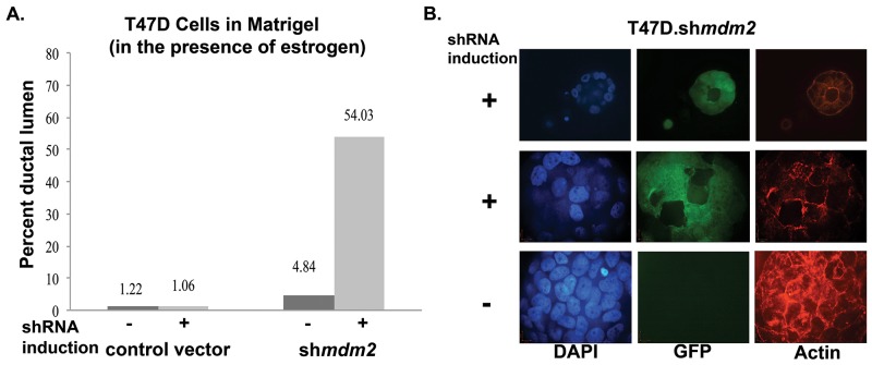
Figure 2. MDM2 depletion in ER+ breast cancer cells with mutant p53 leads to formation of lumen and reverts mammary architecture towards a normal state.
(A) T47D cells grown in matrigel for 3 weeks in presence of estrogen and in the presence or absence of 4 μg/ml dox, were fixed, stained with F-Actin and mounted with DAPI containing mounting media. Confocal z-stack images were acquired. Masses with lumen were counted and presented as percent of total number of masses grown in 3D matrigel. An average of two independent experiments are shown. The number of masses counted for 2 independent experiments were control vector -shRNA induction (21, 41), control vector +shRNA induction (21, 47), mdm2shRNA -shRNA induction (31, 46) and mdm2shRNA +shRNA induction (31, 62). The p-value determined by 2-tailed Student t-test comparing with and without MDM2 knockdown was p-value=0.01. Two independent scorers counted the numbers of masses for each independent experiment. (B) A representative image from confocal immunofluorescence microscopy showing a single slice from z-stack of DAPI, GFP and F-Actin of estrogen treated inducible clonal T47D.shmdm2 cells grown in 3D-matrigel in the presence or absence of 4μg/ml doxycycline (dox) for 3 weeks. The top and middle rows show hollow lumen and ductal lumen respectively in the presence of shRNA expression to mdm2; the GFP (green) indicates shRNA induction to mdm2. The third row shows mass structure (disruption of normal mammary glandular architecture) in the absence of shRNA expression to mdm2.

