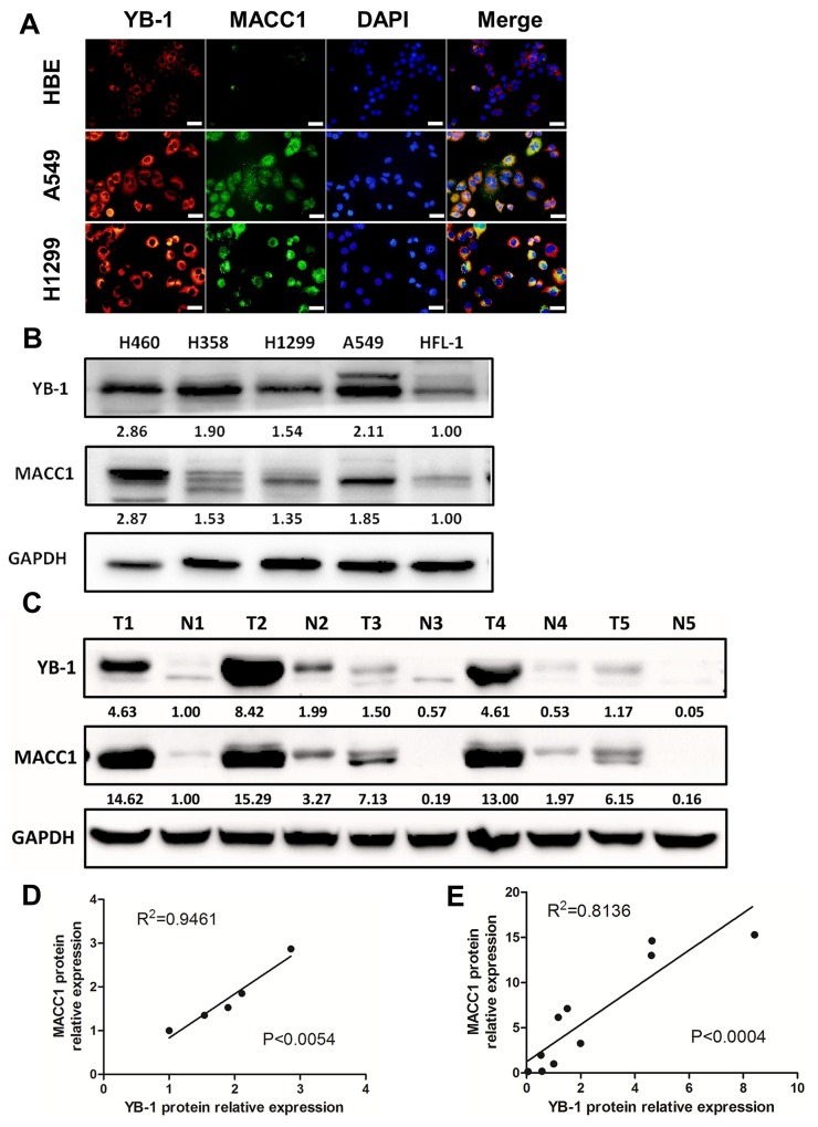
Figure 5. YB-1 was positively correlated with MACC1 and both proteins were over-expressed in lung adenocarcinoma cell lines.
(A) Co-localization of YB-1 and MACC1 in HBE, A549 and H1299 cells by immunofluorescence assay. Red: YB-1; Green: MACC1. Scale bars, 20 μm. Lung adenocarcinoma cell lines (B) and lung adenocarcinoma tissues (C) were lysed and subjected to Western blot to analyze YB-1 expression and MACC1 levels (T, lung adenocarcinoma tumor; N, normal lung tissues adjacent to tumor lesions). YB-1 and MACC1 protein expression levels were normalized to HFL-1 in lung adenocarcinoma cell lines. Meanwhile, those were normalized to N1 in lung adenocarcinoma tissues. Moreover, linear regression analysis was performed, (D) in lung adenocarcinoma ccell lines, (E) in lung adenocarcinoma tissues.

