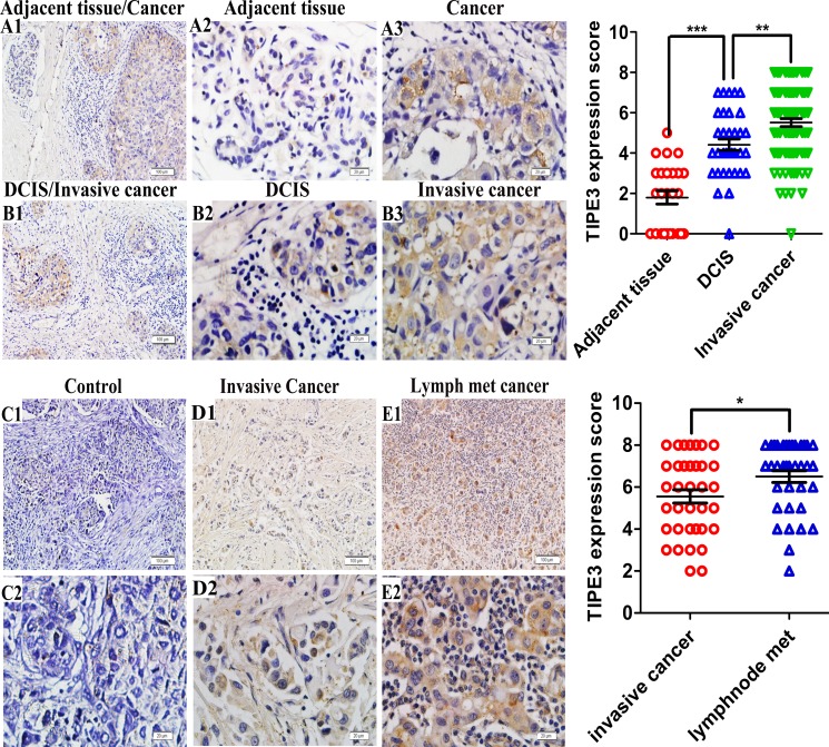
Figure 1. TIPE3 expression in breast cancer as determined by IHC.
A1 and A2 showed TIPE3 expression in adjacent breast tissue (A1, 100 ×; A2, 400 ×). TIPE3 protein was detected in ductal carcinoma in situ (DCIS) and invasive ductal carcinoma (B1, 100 ×; B2, B3 and A3, 400 ×). D1 and D2 showed TIPE3 expression in invasive ductal carcinoma (D1, 100 ×; D2, 400 ×). E1 and E2 showed TIPE3 expression in lymphatic metastasis carcinoma (E1, 100 ×; E2, 400 ×). Isotype control was not staining (C1, 100 ×; C2, 400 ×). All of the experiments were repeated at least three times with similar results and representative data are shown. (*P < 0.005, **P < 0.001 and ***P < 0.001).

