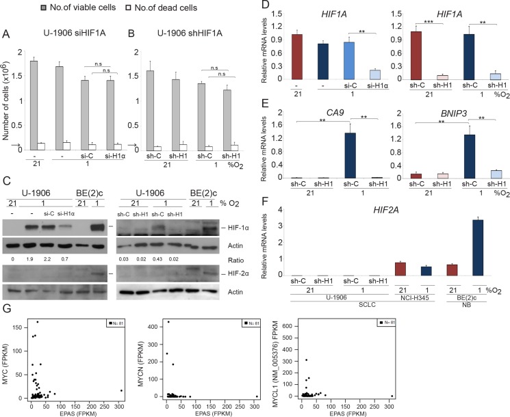
Figure 1. SCLC cells survive and proliferate at 1% oxygen despite HIF1A knockdown.
U-1906 cells were (A) transfected with siRNA (si-H1α) or lipofectamine alone (−) or (B) transduced with shRNA (sh-H1) against HIF1A or a non-targeting control (si-C and sh-C). The cells were cultured for 72 hours at 21% or 1% oxygen. The number of viable and dead cells was counted in triplicates. The arrows indicate number of cells seeded at day 0. (C) HIF-1α and HIF-2α western blot analyses of whole cell lysates. SK-N-BE(2)c neuroblastoma cells (BE(2)c) grown at 1% oxygen for 4 hours and 72 hours were used as positive HIF controls. Actin was used as loading control. The ratio between HIF-1α/Actin was calculated. (D–F) The relative mRNA expression levels of HIF1A, HIF2A, BNIP3 and CA9 were analyzed by qPCR and the expression data were normalized to three reference genes (HPRT1, UBC, TBP). Error bars show the standard deviation and 2-tailed unpaired Student's t test were performed. * indicates p < 0.05, ** < 0.01, *** < 0.001. (G) Expression of MYC (left panel), MYCN (center), and MYCL1 versus EPAS for 81 SCLC tumors reported by George et al. (PMID:26168399).

