
Figure 1.
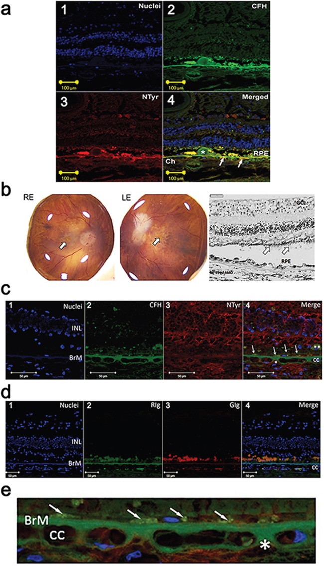
a. Immuno-localisation of CFH and nitrotyrosine in the choroid and retina in an early AMD human eye (1-4). (1) Nuclei are counterstained with bisbenzimide (blue). (2) Double immunolabelling for CFH (green) and (3) nitrotyrosine (NTyr) (red) in the retina and choroid from an eye with early AMD (83 year old, female, AREDS Grade 1). Image in 4 is merged and shows colocalisation (yellow) of CFH and NTyr in Drusen (*), RPE, Bruch's membrane and extracellular matrix around the choriocapillaris (Ch). Arrows indicate site of colocalisation. b. Right (RE) and left (LE) postmortem eyes (93 yo, dry AMD AREDS grade 4) photographs showing areas of atrophy in the macular region of each eye (arrow – fovea). Representative image of a haematoxylin & eosin stained paraffin section of the retina and choroid from the RE dry AMD lesion, showing disrupted retinal pigment epithelium (RPE) and sub-RPE deposits. Note the thinned outer nuclear layer and remnants of photoreceptor outer segments (arrows) (GCL: ganglion cell layer; INL: inner nuclear layer). c. Immuno-localisation of CFH and nitrotyrosine in the choroid and retina in the human eye shown in b. (1) Nuclei are counterstained with bisbenzimide (blue). (2) Immunostaining for CFH (green) and (3) NTyr (red). (4) Colocalisation of CFH and NTyr are seen in the BrM (arrows point to area of colocalisation). Small areas of CFH and NTyr co-localisation (merge, *) are seen in the extracellular matrix around the choriocapillaris (cc). Remnants of RPE are visible (**) in this area, and the outer nuclear layer is not visible, associated with outer retinal atrophy. d. Immunolabelling with immunoglobulin (Ig) control (1) Nuclei are counterstained with bisbenzimide (blue). (2) Rabbit (RIg) (green) and (3) goat (GIg) (red) shows some non-specific immunolabelling (cc: choroiocapillaris; INL: inner nuclear layer; BrM: Bruch's membrane). e. High power image of Figure 1c (4) demonstrating immuno-localisation of CFH (green) and nitrotyrosine (red) in the Bruch's membrane (BrM) / choroiocapillaris region of the dry AMD human eye. Nuclei are counterstained with bisbenzimide (blue). Small areas of CFH and NTyr co-localisation (merge, *) are seen in the extracellular matrix around the choriocapillaris (cc) and in cell remnants just above Bruch's membrane (arrows).