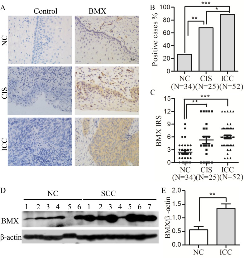
Figure 1. BMX expression is up-regulated in cervical carcinomas.
(A) Immunohistochemistry (IHC) for BMX expression is shown in the normal human cervix (NC, n = 34), cervical carcinoma in situ (CIS, n = 25) and invasive cervical carcinoma (ICC, n = 52); scale bar is 10 μm. (B) Analysis of the percentage of BMX-positive cells in NC, CIS and ICC using a chi-square test. (C) The average immunoreactivity score (IRS) of BMX staining in NC, CIS and ICC; one-way ANOVA was performed. (D) Western blot analysis of BMX expression in normal cervix (NC, n = 6) and invasive cervical carcinoma (ICC, n = 7) is shown. (E) The relative quantitative analysis of BMX expression according to western blot results using Quantity One software; a t-test was performed. Values are shown as the mean ± SEM, *p < 0.05, **p < 0.01, and ***p < 0.001.

