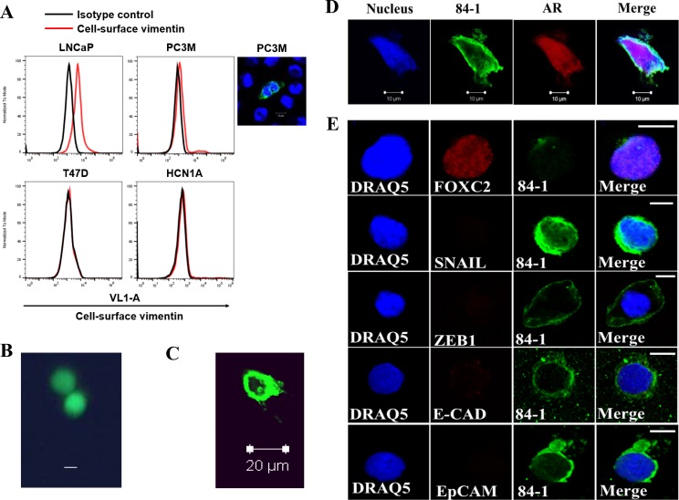
Figure 1. Detection of CSV-CTCs from patients with metastatic prostate cancer.
(A) Immunological assessment of CSV expression in CSV-expressing LNCaP, PC3M (cell staining shown adjacently), and CSV-nul T47D and HCN1A cell lines using flow cytometry. CSV was detected by 84-1 antibody (red line). Isotype control was used as negative control (black line). (B, C) Spiked Calcein AM stained LNCaP cells were isolated using CSV method and analyzed under fluorescent microscope, Scale bar, 10 μm (B) and confocal microscope (C). (D) CTC isolated from patient blood was stained for androgen receptor (AR) and CSV using 84–1. Scale bar, 10 μm. (E) CTCs stained with CSV and various markers as indicated. All experiments were done in triplicates.

