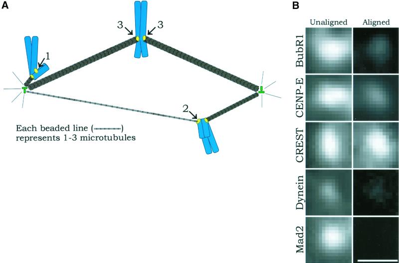
Figure 4.
(A) Diagram showing relative numbers of kinetochore microtubules for unattached kinetochores on mono-oriented chromosomes (1; no kinetochore microtubules), leading kinetochores on congressing chromosomes (2; according to McEwen et al., 1997, these generally possess a few microtubules and can be grouped with unattached kinetochores), and fully attached kinetochores on chromosomes aligned at the metaphase plate (3; these are bound to mature kinetochore fibers, which contain ∼25 microtubules [McEwen et al., 1997]) in prometaphase cells. Nonkinetochore spindle microtubules are not shown for clarity. (B) Immunofluorescence images comparing the fluorescence intensity of unattached or leading kinetochores of unaligned chromosomes and fully attached kinetochores of metaphase-aligned chromosomes for the proteins Bub1R, CENP-E, CREST, cytoplasmic dynein, and Mad2. CREST remains unchanged, BubR1 and CENP-E deplete to moderate levels, cytoplasmic dynein depletes to a low level, and Mad2 becomes undetectable. The pixel density in the photographic images is higher than in the original micrographs. Scale = 1 μm.

