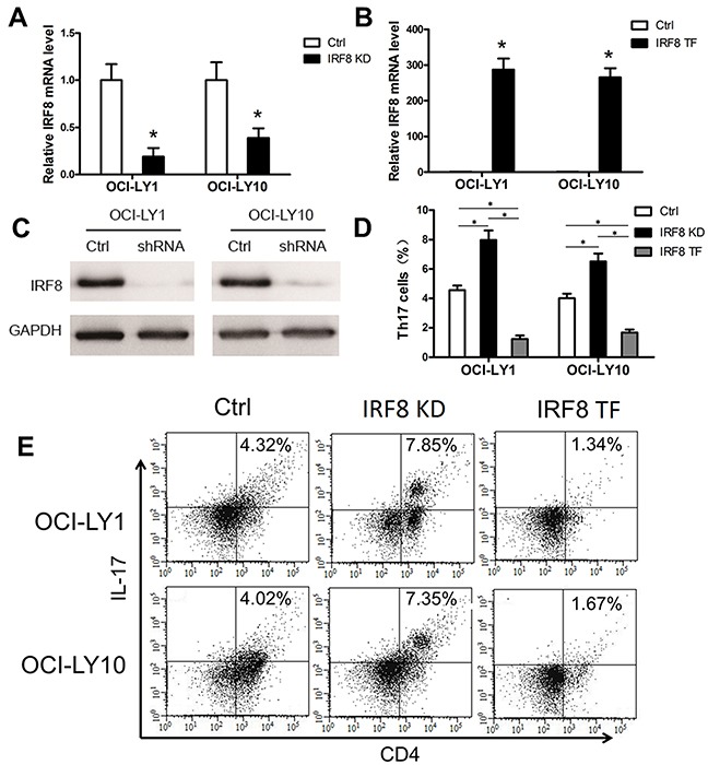
Figure 5. The decline of IRF8 in DLBCL cell lines promoted the generation of Th17 cells and the increase of IRF8 inhibited it in vitro.

One shRNA clone was used to knock down the level of IRF8 in the DLBCL cell lines OCI-LY1 and OCI-LY10. The level of IRF8 transcripts (A) and IRF8 proteins (C) decreased in OCI-LY1 and OCI-LY10 with IRF8 knockdown (IRF8 KD). (B) OCL-LY1 and OCL-LY10 were transfected with lentiviruses encoding IRF8 (IRF8 TF); the transfected cells expressed high levels of IRF8 mRNA. (D) The percentage of Th17 cells from the IRF8 KD group was significantly higher than that from the Ctrl group; the IRF8 TF group was significantly lower than the Ctrl group and the IRF8 KD group. (E) FACS plots of Th17 cell percentages in PBMCs cultured in vitro. Data represent one of three independent experiments. PBMCs from healthy donors were co-cultured with OCI-LY1 and OCI-LY10 (Ctrl), IRF8 KD, or IRF8 TF. All three groups were co-cultured with IL-2, TGF-β and IL-1β for 7 days and then detected by FACS. Error bars represent SD. Significance was determined using independent-sample Student's t/t′ test (two groups) or single-factor analysis of variance (one-way ANOVA) Student-Newman-Keulor/Dunnett's T3 test (three groups). (*, P<0.05).
