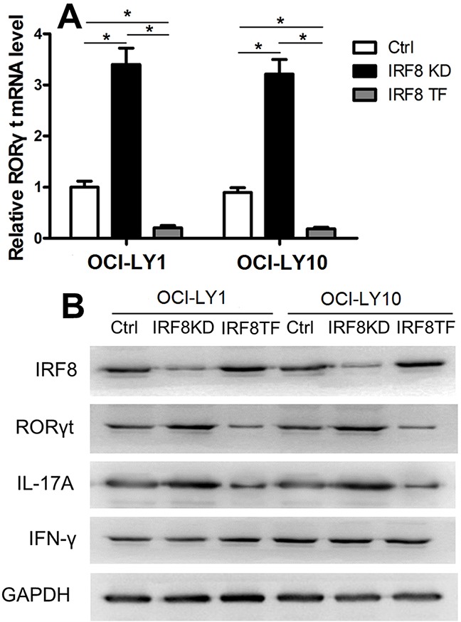
Figure 6. IRF8 suppressed RORγt expression in CD4+T cells.

(A) The RORγt mRNA level was significantly higher in the IRF8 KD group and lower in the IRF8 TF group compared with the Ctrl group. (B) Western blot analysis of the Ctrl, IRF8 KD, and IRF8 TF groups in CD4+T cells using specific antibodies against IRF8, RORγt, IL-17, IFN-γ, and GAPDH. GAPDH was used as a loading control. Error bars represent SD. Significance was determined using single-factor analysis of variance (one-way ANOVA) Student-Newman-Keulor/Dunnett's T3 test. (*, P<0.05).
