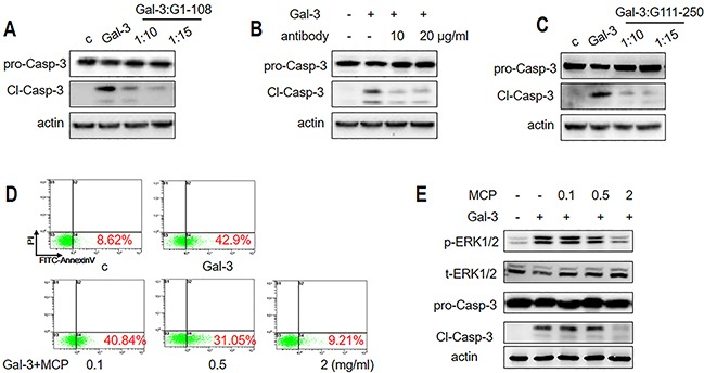
Figure 7. Gal-3-induced T cell apoptosis is inhibited by NT or CRD inhibitors.

Jurkat cells were incubated with 1 μM Gal-3 for 18 h in the presence or absence of (A) 10 or 15 μM G1-108 variant, (B) 10 or 20 μg/ml A3A12 antibody or (C) 10 or 15 μM G111-250 variant. Caspase-3 cleavage was assessed by western blot. (D-E) Jurkat cells were treated with 2.5 μM Gal-3 in the absence or presence of 0.1, 0.5 and 2 mg/ml MCP for 18 h. The apoptotic rate was measured by flow cytometry (D), and p-ERK1/2 and cleaved caspase-3 were determined by western blotting (E). The data are representative of three independent experiments.
