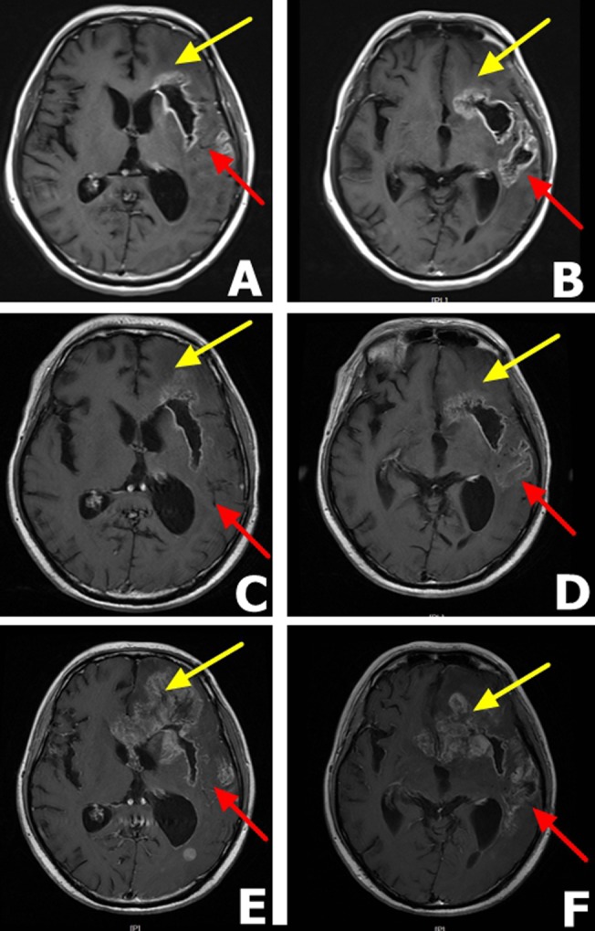
Figure 4. MRI findings in a male patient presenting with glioblastoma multiforme after erlotinib 150 mg therapy A. and B. MRI scans after 14 days of daily erlotinib 150 mg therapy.

The scans reveal tumor hollowing, lower grade edema and reduced compression of the lateral ventricles. C. and D. Three months after erlotinib 150 mg therapy, MRI scans shows that the tumor stabilized. Yellow arrow shows a stable frontal lobe while the red arrow show partial response in the temporal lobe. E. and F. Five months after erlotinib 150 mg therapy. MRI scans demonstrate the tumor relapsed for the second time with distant metastasis and the patient died two months later. The yellow arrow shows obvious metastasis in the frontal lobe and red arrow shows a stable temporal lobe.
