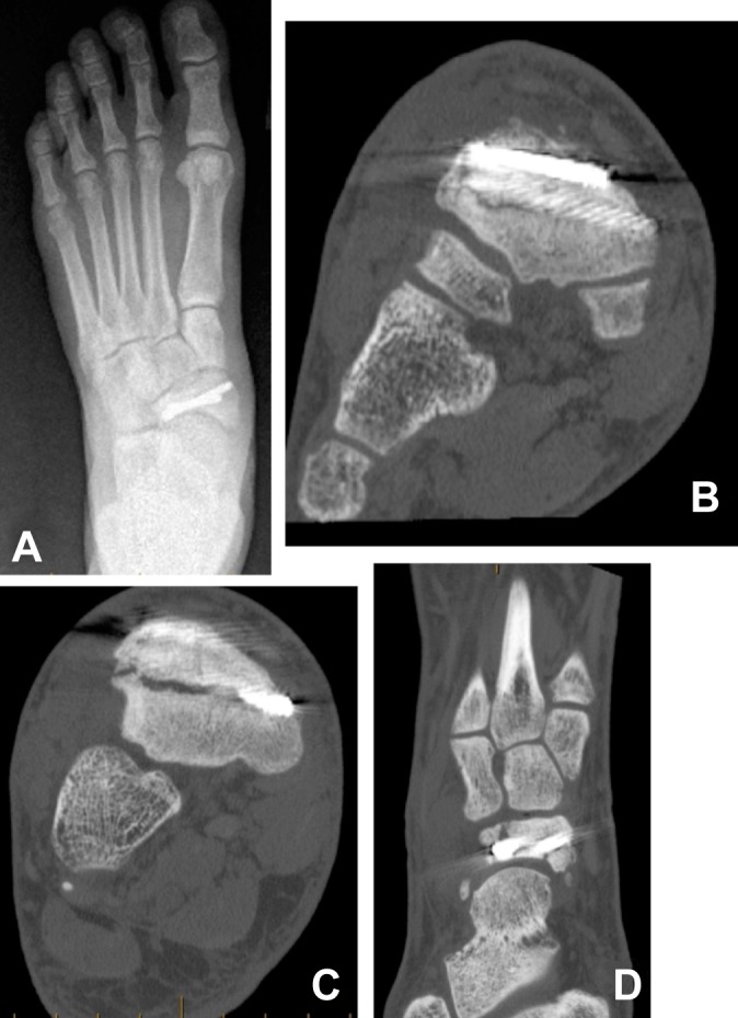
Figure 2.

(A) Anteroposterior radiograph of a player’s right foot after 2 screws were placed lateral to medial. (B) The coronal computed tomography scan demonstrates that the fracture is still present. The (C) coronal and (D) axial cuts of the scan, 2 months later, demonstrate a refracture around the screws.
