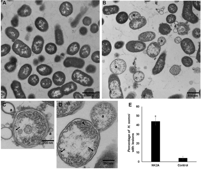
Fig 6. NK2A-induced damage to H. somni cell membranes.
(A) Control H. somni. (B), (C), and (D) H. somni were incubated with 30 μM NK2A peptide at 37°C for 60 min. Arrows indicate damaged inner membranes and letter “a” indicates protrusions on outer membrane. (E) Percentage of H. somni with membrane damage. Means and standard deviations were calculated from six electron micrographic images of control and NK2A treated samples. (P<0.001)

