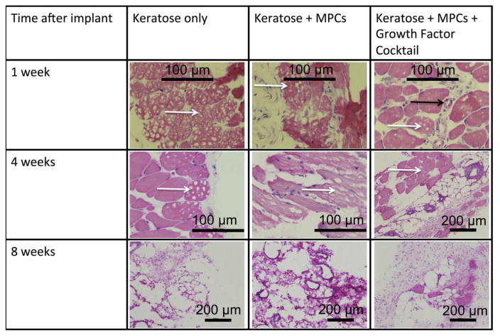
Figure 7. H&E images of explanted keratose hydrogels.
Keratose hydrogels were removed at 1, 4, and 8 weeks. Representative H&E images from the keratose only, keratose loaded with MPCs, and keratose loaded with MPCs and growth factors. There were no signs of inflammation. Cells were observed within the keratin hydrogel at 1 and 4 weeks that were consistent with blood vessel and muscle progenitor cells. At the 8 week time point the keratose only group had degraded and was replace with morphology consistent with normal tissue in the subcutaneous space, while the cell loaded keratose was replaced with a highly vascularized, mix of cell consistent with the morphology of muscle progenitor cells. Black arrow indicates location of a blood vessel while white arrows indicate the presence of keratose gel remaining at the implant site. Scalebars are as indicated in the figure.

