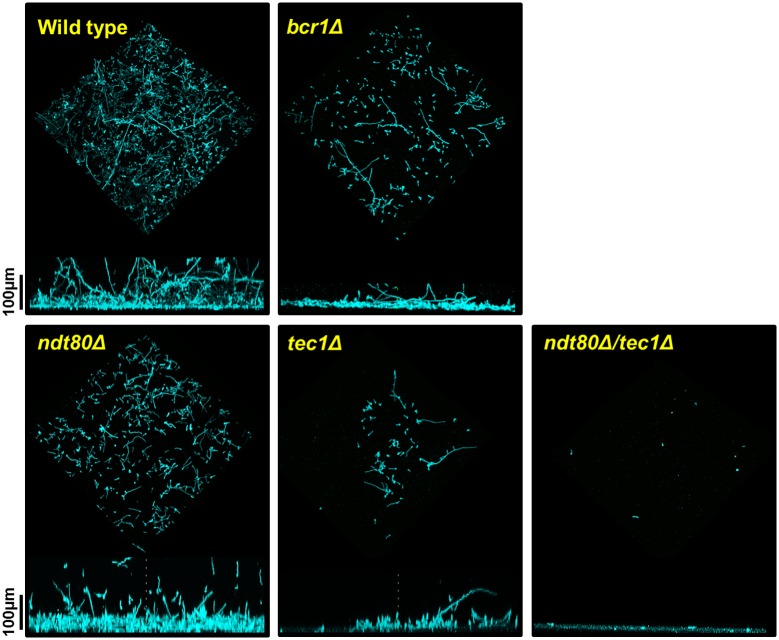
Fig 4. The ndt80Δ/NDT80 and bcr1Δ/BCR1 show altered biofilm architecture.
Confocal microscopy images of wild type, ndt80Δ/NDT80, bcr1Δ/BCR1, tec1Δ/TEC1, and ndt80Δ/NDT80 tec1Δ/TEC1 strains grown in YETS for 48 hr. Cells were stained with concanavalin A. See Materials and methods for full details. The top image is a three-dimensional rendering of the biofilm. The bottom image is a cross-sectional view of the same biofilm.

