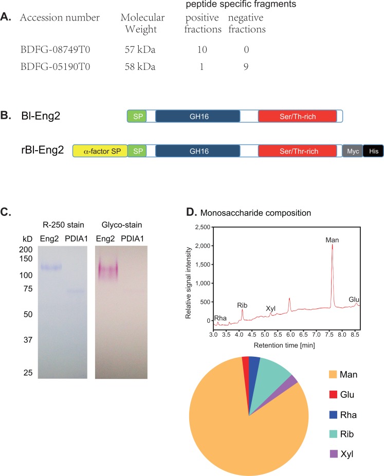
Fig 2. Mass spec analysis identified Bl-Eng2 as a Dectin-2 ligand candidate.
(A) The ligand- negative and -positive fractions (#9–13 and #1–7 from S1C Fig, respectively) from the second gel filtration were analyzed by Mass spectrometry. Numbers on the right represent number of peptide specific fragments detected. (B) Domains of native B. dermatitidis Eng2 (Bl-Eng2) and recombinant Bl-Eng2 expressed in Pichia pastoris: SP denotes Signal peptide; GH16 denotes glycosyl hydrolase catalytic domain; Ser/Thr-rich domain harbors 68 potential O-linked glycosylation sites; and Myc and His tags are placed at the C terminus for purification. (C) 0.6 μg Bl-Eng2 and 0.3 μg PDIA1 were run on SDS-PAGE gel under reducing conditions and stained for protein (left) or carbohydrate (right). (D) Monosaccharide composition of Bl-Eng2 measured by gas chromatography (GC). GC chromatogram of the alditol acetate-derivatized monosugars of hydrolyzed Bl-Eng2 (top). Monosaccharides are labeled as follows: Rha—rhamnose, Rib—ribose, Xyl—xylose, Man—mannose, and Glu–glucose. Unlabeled peak at 5.953 min resulted from component degradation during alditol acetate derivatization. Pie diagram shows the relative contribution of monosaccharides (bottom).

