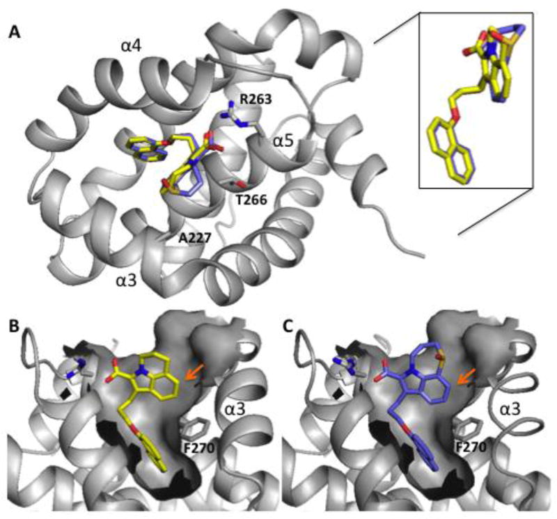
Figure 2.
X-ray structures of tricyclic indole acids bound to Mcl-1. (A) Compounds 17 and 24 interact with Mcl-1 in the BH3-peptide binding cleft between helices 3, 4, and 5. The indole cores and naphthyl groups adopt almost the same binding poses while C-ring units adopt different binding conformations (inset). Shown is the surface depiction of Mcl-1 when complexed to (B) Compound 17 and (C) Compound 24 that illustrates how these ligands occupy the P2 pocket. Arrows indicate potential positions for substitutions to fill unoccupied sub-sites.

