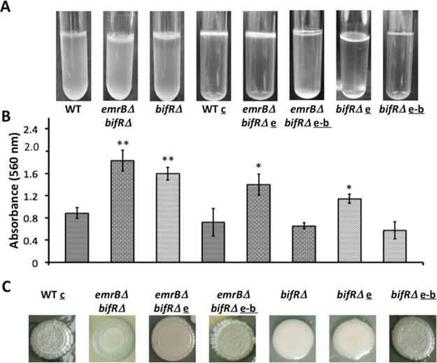
Figure 7.

Deletion of bif R leads to increased biofilm formation. (A) Pellicle formation in static cultures. (B) Quantitation of biofilm using crystal violet staining; corresponding pictures of pellicle formation and strain identification are directly above each bar. Error bars represent standard deviation from three separate cultures. (C) Colony morphology of WT, bif RΔ, emrBΔ-bif RΔ, and corresponding complemented strains. WT and mutant strains were complemented with empty pBBR-MCS5 (denoted c, for WT); e, pBBR-MCS5 encoding emrB; e-b, pBBR-MCS5 encoding emrB-bif R. Asterisks represent statistical significance from WT based on Student’s t test (**, p < 0.001 and *, p < 0.05).
