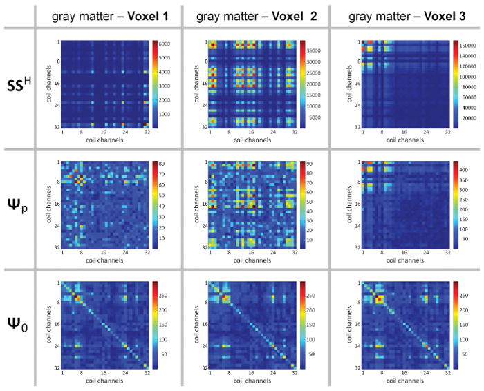
Figure 8.

Varying levels of agreement between the signal and noise statistics. Statistical matrices are shown across coil channels taken from a single representative subject (1×1×3 mm3 images, 3T, 32Ch array coil). (Top row) Average (over the time-series) of the signal level matrix SSH. (Middle row) Physiological noise covariance matrix Ψp. (Bottom row) Thermal noise covariance matrix Ψ0. Each column shows the matrixes from a single voxel within the cortical gray matter mask. For Voxel 1, the structure of the SSH matrix is substantially different from the Ψp matrix, whereas for Voxel 2 there is some common structure between these two matrices, while for Voxel 3 the two matrices appear to be identical up to a global scaling. Note that the Ψ0 matrix is virtually identical across the voxel positions, and is distinct from both the SSH and Ψp matrices.
