
Fig. 2. In vitro axotomy recapitulates the phenotypic strain differences in regeneration observed after in vivo injury.
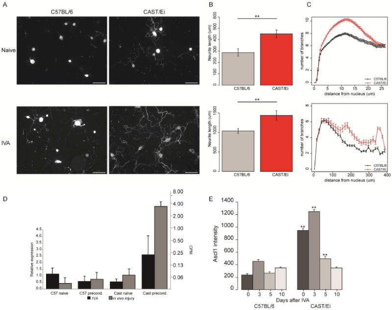
(A) Representative fluorescence images of C57BL/6 and CAST/Ei DRG neurons stained against TUBB3 highlighting neurite outgrowth in both strains with or without an IVA. [Scale bar = 100 μm] (B-C) Quantification of neurite length (B) and complexity (C) in the naïve (upper panel) and IVA (lower panel) C57BL/6 (black) and CAST/Ei (grey) neurons. The CAST/Ei neurites are longer and more complex with a greater arborization, in both conditions. (D) IVA (black) recapitulates the increase in Ascl1 mRNA levels observed after in vivo injury (grey). (E) Time course of Ascl1 protein expression levels in C57BL/6 and CAST/Ei neurons using immunocytochemistry. Ascl1 levels were higher up to 5 days after IVA, in the CAST/Ei neurons than C57BL/6 neurons. By day 10, the levels were not different from baseline in both strains after IVA. (**p≤1e-2, Student’s t-test).