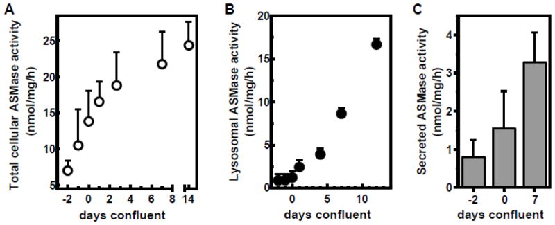
Figure 1. ASMase activity elevates over time in cultured endothelial cells.
A: BAEC were harvested at different days after seeding as indicated and ASMase activity was determined in cell lysates using bovine [14C-methylcholine]sphingomyelin. Confluence was judged visually using a phase-contrast microscope (day 0 was defined as the day the monolayer covered 100% of the surface of the well). B: ASMase activity was determined in absence of exogenous zinc, quantifying the lysosomal fraction of cellular ASMase. C: BAEC culture medium (serum-free) was collected after 24 hours and concentrated using 30 kDa molecular weight cut-off filter units. ASMase activity elevates over time in concordance with total cellular levels. Data (mean ± SD) are collated from 9 separate experiments.

