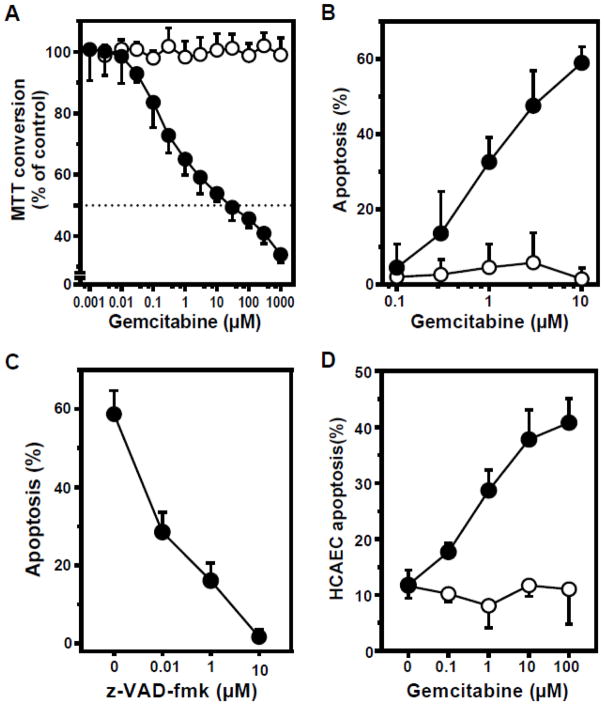
Figure 5. Proliferating endothelial cells are gemcitabine sensitive, in contrast to growth-arrested cells.
Endothelial cells were either proliferating (closed circles), or confluent at growth-arrest (open circles). A: BAEC were exposed to gemcitabine and MTT conversion was measured as a readout for cell viability. Cells treated with vehicle as a control were set at 100%. B: After 16 hours gemcitabine exposure, BAEC cells were harvested and apoptosis was quantified by bis-benzimide nuclear staining. C: Pre-treatment (45 min) of BAEC with the pan-caspase inhibitor z-VAD-fmk blocks apoptosis induced by 10 μM gemcitabine. D: HCAEC were exposed to gemcitabine for 16 hours and apoptosis was quantified. Data (mean ± SD) are collated from 3 separate experiments each.

