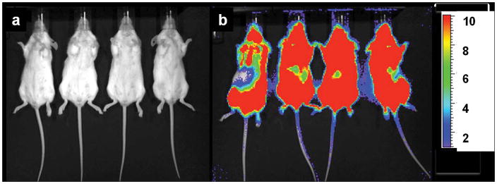
Figure 2.
Bioluminescence imaging (BLI) of two groups of scid mice that were xenografted with Daudi tumor cells transfected with the green fluorescent protein (GFP) and firefly luciferase (FFLuc) genes [45]. Images were taken on day 17 (a) after treatment with [225Ac]DOTA-B4 or (b) untreated growth controls. In the scid model, the GFP+/FFLuc+ Daudi cells developed into macroscopic, disseminated tumors in the bone marrow and spleen as well as in kidneys, liver, lungs, ovaries, and adipose tissue. BLI clearly showed the presence of lymphoma in the untreated mice while no disease was detected in the mice treated with the [225Ac]DOTA-B4. (n.b., the scale bar indicate the value x 1E6 photons/sec/cm2).

