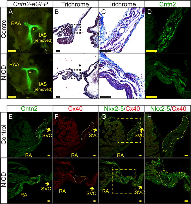
Figure 2. Nodal structure is preserved with mis-expression of Nkx2-5 in iNICD nodal myocardium.

(A) Whole-mount pictures of the RA endocardial surface from a representative control (top, αMHC-rtTA; Cntn2-eGFP) and iNICD (bottom, αMHC-rtTA; tetO_NICD; Cntn2-eGFP) heart indicate no gross difference in Cntn2-eGFP expression delineating overall conduction system morphology (Scale bars=125 μM). Mice were between 5–6 months of age at the start of Notch induction for 3 weeks, followed by a 5-month washout. (B) Representative Masson’s Trichrome images from a control (top, αMHC-rtTA) and iNICD (bottom, αMHC-rtTA; tetO_NICD) SAN located at the junction of the SVC with the RA. Boxed regions in B (scale bars=100 μM) are enlarged in Panel C (scale bars=50 μM). (C) No excess fibrosis was detected within the iNICD SAN to account for the observed sinus bradycardia. (D) Serial sections to Panel C stained for Cntn2 further delineate the region of the SAN (scale bar=50 μM). (E,F) The SAN is histologically identified as Cntn2+ (E) and Cx40− (F) myocardium at the junction of the SVC and RA. Yellow dashed lines demarcate the compact SAN, which has a similar morphology between control (Supplemental Figure V) and iNICD mice (Supplemental Figure VI). Scale bars E,F=20 μM. (G,H) Nkx2-5 is mis-expressed within the compact SAN of iNICD mice. Yellow boxed region in G (scale bar=20 μM) is enlarged in Panel H (scale bar=50 μM) to better visualize the SAN. Mice were started on doxycycline chow between 2–3 months of age and remained on doxycycline chow for 4 weeks with no washout. IAS = inter-atrial septum, RAA = right atrial appendage, SVC = superior vena cava, RA = right atrium. * indicates lumen of SVC near region of the SAN.
