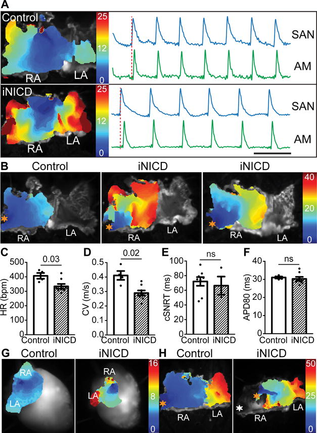
Figure 4. Transient Notch activation results in stable slowing of atrial conduction velocity.

(A) Activation maps from isolated atria from a control and iNICD heart during sinus rhythm demonstrate that, despite a slower HR, the location of the dominant pacemaker remains at the junction of the SVC and RA in iNICD hearts. Optical action potentials of the SAN region and distant atrial myocardium demonstrate 1:1 activation in both control and iNICD atria. Together with RR interval plots of iNICD mice demonstrating that the pauses are not multiples of the preceding RR interval (Supplemental Figure X), the observed bradycardia is not likely due to exit block. Scale bar=0.2 seconds. (B) Activation maps of the RA obtained by pacing the RA appendage (orange asterisk) demonstrate overall slower conduction in iNICD hearts when compared with control. In addition, isochrones crowding and focal areas of conduction block were observed in iNICD atria. Under Langendorff perfusion, iNICD hearts exhibit slower HR (C). Optical mapping of isolated atria showed slower CV (D) in iNICD mice when compared with controls while the corrected SAN recovery time (cSNRT) (E) and APD80 (F) remain unchanged. In A–F, mice were fed doxycycline chow at 2 months of age for 3 weeks, followed by a washout period of 2–3 months. iNICD mice were αMHC-rtTA; tetO_NICD (n=4 females, n=4 males) and controls were littermate tetO_NICD (n=4 males). (G) Notch induction in juvenile mice (3 weeks of age) results in regional atrial inexcitability as evidenced by the absence of depolarization in a portion of the RA during spontaneous sinus rhythm (left), and an inability to capture with RA appendage pacing (H, white asterisk, right). iNICD were αMHC-rtTA; tetO_NICD and controls were littermate tetO_NICD. Statistics were performed using unpaired t tests with Welch’s correction. Values of P<0.05 were considered statistically significant. RA = right atrium, LA = left atrium, SAN = sinoatrial node, AM = atrial myocardium, ns = no statistical difference.
