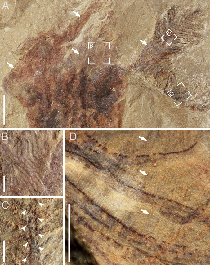
Fig. 2.
Tentacle architecture of X. sinica (ELEL-SJ080827B; SI Appendix, Fig. S1). (A) Upper part showing numerous pinnate tentacles (arrowed). (B–D) Close-up of focus areas in A showing sinuous pinnules (B), attachment sites (arrowheads) of pinnules branching from the rachis (C), and closely spaced cilia (arrows) fringing the pinnules (D), respectively. [Scale bars: 10 mm (A), 1 mm (B–D).]

