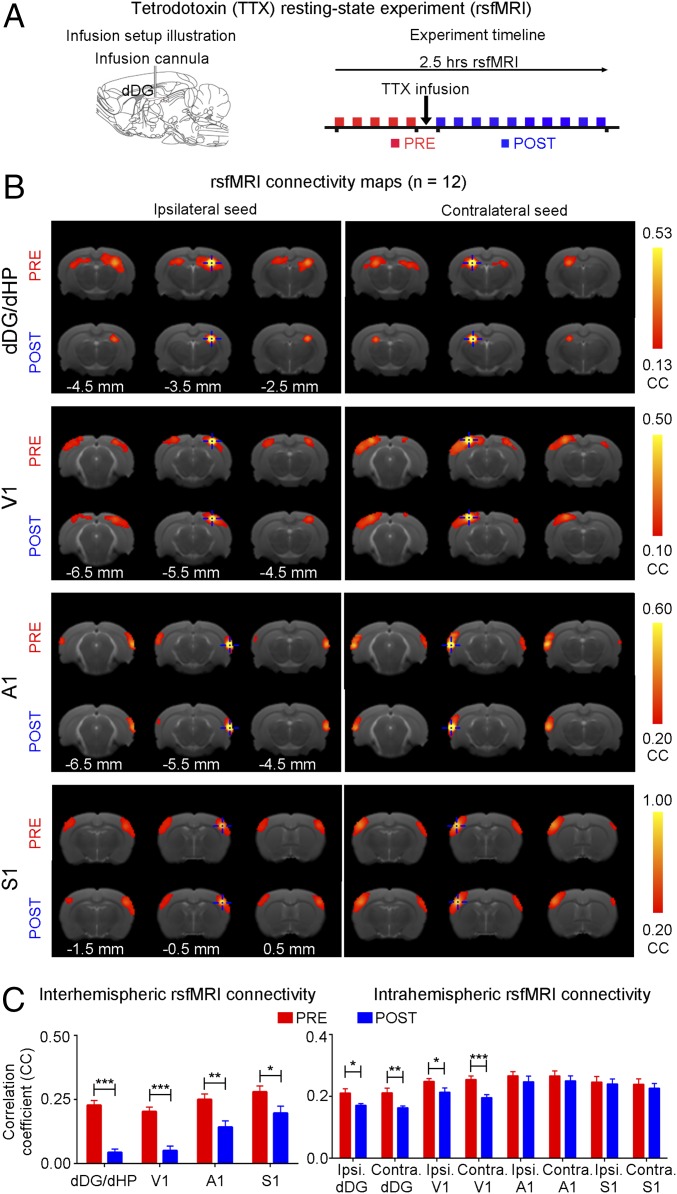
Fig. 7.
Pharmacological inactivation of dDG neurons in dHP decreases interhemispheric rsfMRI connectivity. (A) Illustration of the tetrodotoxin (TTX) infusion setup (Left) and a typical rsfMRI experiment timeline whereby TTX is infused into dHP a minute after the last baseline (pre) scan (Right). The first post scan is acquired a minute after the completion of TTX infusion. (B) rsfMRI connectivity maps of dHP, V1, A1, and S1, pre and post infusion of TTX. (C) Quantification of the interhemispheric rsfMRI connectivity (Left; n = 12; paired t test followed by Bonferroni’s post hoc test; *P < 0.05, **P < 0.01, and ***P < 0.001; error bars indicate ±SEM).

