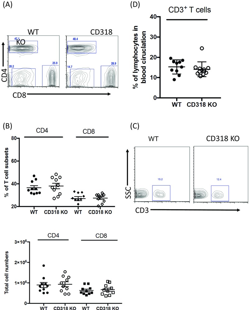
Fig. S1.
Intact T-cell compartment in the lymph node and peripheral blood of CD318 KO mice. (A) A representative frequency of CD4+ and CD8+ T cells in WT and CD318 KO inguinal lymph nodes. Data are representative of 10 mice. (B) The summary of the frequency and total cell numbers of CD4+ and CD8+ T cells in WT and CD318 KO inguinal lymph node. n = 10 in each group. (C) A representative frequency of CD3+ T cells in WT and CD318 KO peripheral blood. (D) The summary of frequency of CD3+ T cells in the peripheral blood of WT and CD318 KO mice. n = 10 in each group.

