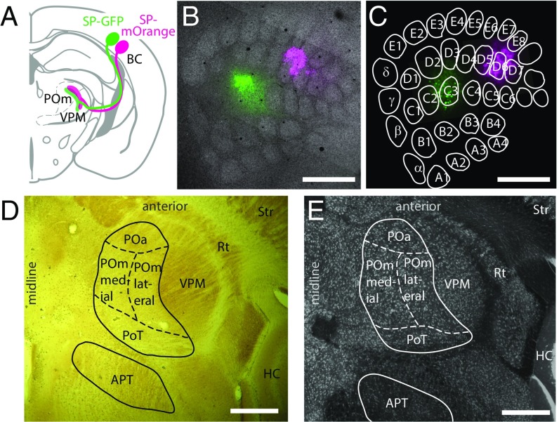
Fig. 1.
Cortical bouton labeling and borders of subcortical target nuclei. Subcortical L5B boutons in the thalamus were labeled by virus-mediated expression of two different fluorescent proteins in barrel cortical neurons. (A) Schematic showing dual injection of viral particles encoding Synaptophysin–GFP (SP–GFP, green) and Synaptophysin–mOrange (SP–mOrange, magenta) into the BC and projections in the thalamus (modified from ref. 12). (B) Merged confocal fluorescence image of tangential sections of the BC at the level of layer 4, showing barrels (gray; Streptavidin staining) and deposits of SP–GFP (green) and SP–mOrange (magenta). (C) As in B, but at the L5B level with labeled L5B somata. Barrel outlines are from B. (D) Horizontal section (cytochrome c oxidase staining) containing L5B target nuclei in the thalamus: (POa, POmmedial, POmlateral, PoT, and APT). The VPM, thalamic reticular nucleus (Rt), striatum (Str), and hippocampus (HC) are indicated for orientation. Zona incerta is shown in Fig. S1. (E) An image at a level similar to that shown in D but stained for neuronal somata by NeuroTrace. (Scale bars: 500 µm in B–E.)

