Fig. 2.
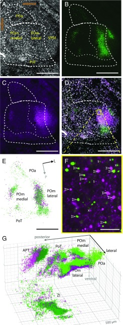
Reconstructions of subcortical L5B bouton clouds. Shown are examples of confocal sections and bouton reconstructions. (A–C) Individual channels of horizontal sections through L5B target nuclei in the dorsal thalamus. (A) Somata (NeuroTrace staining; gray). (B) GFP-labeled L5B boutons (green). (C) mOrange-labeled L5B boutons (magenta). (D) Overlaid sections A–C. Dashed lines indicate borders of PO nuclei. (E) Reconstructed boutons from B and C (green and magenta, respectively). Bouton diameters are up-scaled twofold to increase visibility. Arrows indicate orientation (L, lateral; P, posterior). (F) Enlarged view of the boxed area in D but without the NeuroTrace signal. Arrowheads mark examples of putative giant L5B boutons (>1.5 µm). Small puncta (s, <1.5 µm) were excluded; “x” indicates examples of boutons that had their maximum brightness in an adjacent z-section. (G) 3D illustration of reconstructed boutons from experiments in A–F. Major grid lines are spaced by 500 µm. (Scale bars: 500 µm in A–D and F; 25 µm in E.)

