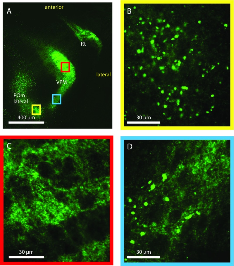
Fig. S3.
Comparison of small L6 and large L5B boutons in thalamus. Micrographs show boutons of different sizes in the VPM and POmlateral. (A) Horizontal overview of a maximal intensity projection over eight slices showing the POmlateral, VPM, and Rt. (B) A single z-section image of the POmlateral from the yellow boxed region in A, showing many large boutons of L5B origin. The POmlateral contains both large L5B and small L6 boutons. (C) A single z-section image of the VPM core area from the red boxed region in A showing a meshwork of small boutons of L6 origin. (D) A single z-section image of the posterior VPM from the blue boxed region in A showing some large boutons of L5B origin amid a meshwork of small L6 boutons. The VPM contains mostly small and dense L6 boutons (C) but also contains some large L5B boutons at the posterior end (D); in the Rt there are only small L6 boutons.

