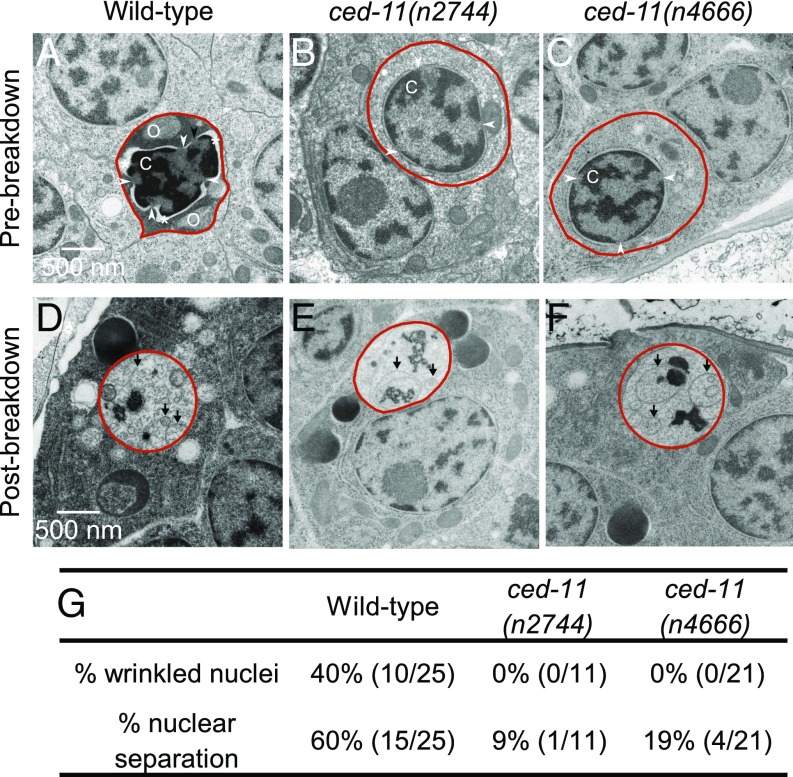
Fig. 2.
Apoptotic cells in ced-11 animals have altered ultrastructure. (A–F) Electron micrographs of engulfed apoptotic cells (outlined in red). (A) A prebreakdown apoptotic cell in a wild-type embryo stained darkly and had a wrinkled nucleus and separation of the nuclear membrane (indicated by the asterisk). The arrowheads indicate the nuclear pore junction. C, condensed chromatin; O, bloated organelle. (B and C) Prebreakdown apoptotic cells in ced-11(n2744) (B) and ced-11(n4666) (C) embryos did not stain darkly and were not distinct from the surrounding living cells. (D–F) Apoptotic corpses postbreakdown in wild-type (D), ced-11(n2744) (E), and ced-11(n4666) (F) embryos contained membranous whorls and did not stain darkly. Arrows indicate a membranous whorl. (G) Percent of prebreakdown apoptotic cells in wild-type and ced-11 embryos with wrinkled nuclei and nuclear separation.

