Fig. 3.
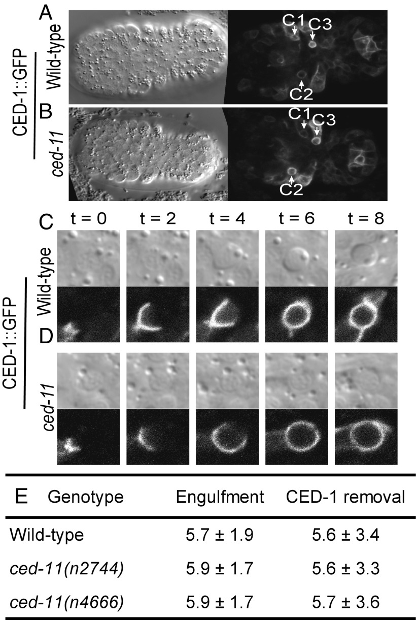
Apoptotic cells in ced-11 embryos are engulfed normally. (A and B) DIC (single focal plane, so not all corpses can be seen) and CED-1::GFP (merge of multiple focal planes) images of wild-type (A) and ced-11 (B) embryos at the onset of ventral enclosure when apoptotic cells C1, C2, and C3 have been engulfed. All strains contain enIs7 (Pced-1::ced-1::gfp). (C and D) CED-1::GFP clustering around apoptotic cells as they are engulfed in wild-type (C) and ced-11 (D) embryos with accompanying DIC images. Images were recorded every 2 min; time 0 is determined by the time of the image taken before CED-1::GFP began to encircle the apoptotic cell. Cells are engulfed at 6 min. (E) Average times for CED-1::GFP accumulation (engulfment) and removal (phagosome maturation) in wild-type and ced-11 embryos. At least nine embryos and 26 apoptotic cells were analyzed for each genotype. Mean time is shown in minutes ± SD.

