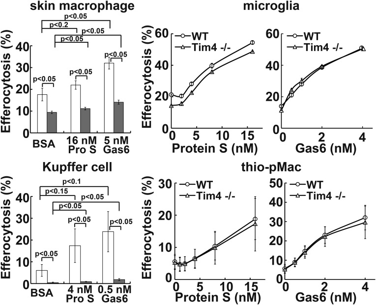
Fig. 6.
Different requirement of Tim4 for efferocytosis by tissue macrophages. The indicated tissue macrophages from wild-type (open column) or Tim4−/− (closed column) mice were seeded into 24-well plates, and the adherent cells were coincubated with pHrodo-apoptotic thymocytes at 37 °C for 1 h (thioglycollate elicited peritoneal macrophages, thio-pMac) or 2 h (others) in DMEM containing 5 mg/mL BSA and the indicated concentrations of mProS or mGas6. Cells were detached and stained with PE/Cy7-anti-CD11b and AlexaFluor647-anti-CD169 (skin macrophages), APC-anti-F4/80 (Kupffer cells), or APC-anti-CD11b (thio-pMac). The percentage of CD11b+CD169+ cells (skin macrophages), F4/80high and autofluorescence-positive cells (Kupffer cells), or CD11b+ cells (thioglycollate elicited peritoneal macrophages) that was pHrodo+ was determined by flow cytometry. For microglia, the percentage of pHrodo+ cells was determined for the total cell population. Experiments were performed in triplicate for skin macrophages and primary microglia or independently three times for Kupffer cells and thioglycollate-elicited peritoneal macrophages, and the average value was plotted with SD (bar) as efferocytosis. P values are shown.

