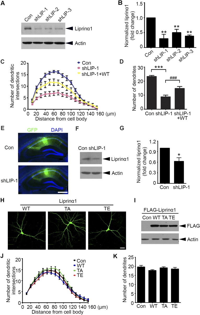
Fig. S2.
Liprinα1, but not its phosphorylation status, is required for dendrite maintenance. (A and B) Western blot analysis of liprinα1 expression in cultured cortical neurons transfected with pSUPER vector as control (Con) and liprinα1 shRNAs. Protein level normalized to actin; **P < 0.01 vs. Con, Student’s t test; n = 3 independent experiments. (C and D) Liprinα1 knockdown resulted in significant dendritic defects, which were partially rescued by an RNAi-resistant human liprinα1 construct (WT). Cultured hippocampal neurons were transfected with the indicated plasmids at 12 DIV and cultured for 5 d. (C) The dendritic complexity of transfected neurons was analyzed by Sholl analysis. Knockdown of liprinα1 reduced the dendritic intersections 10–120 µm from somas compared with Con, which was partially rescued by WT. (D) Quantification of dendrites of transfected neurons. ***P < 0.001 vs. Con; ###P < 0.001 vs. shLIP-1, one-way ANOVA with the Student–Newman–Keuls test; n = 14, 12, and 12 neurons for Con, shLIP-1, and shLIP-1+WT, respectively. (E–G) Liprinα1 expression was significantly reduced in the hippocampal CA1 region by lentiviral infection. (E) Representative images of mouse hippocampal slices infected by lentivirus containing shLIP-1 or GFP control (Con). Slices were stained with GFP (green) and DAPI (blue). (Scale bar: 500 µm.) (F) GFP-infected CA1 regions were dissected for Western blot analysis. (G) Quantification of liprinα1 expression. Normalized to actin; *P < 0.05, Student’s t test; n = 4 mice for each condition. (H and I) The expression levels of different liprinα1 constructs are comparable. (H) Immunostaining of overexpressed liprinα1 (green) in cultured neurons. (Scale bar: 25 μm.) (I) Western blot analysis of the overexpression levels of liprinα1 and its mutants in HEK293T cells. (J and K) Overexpression of TA or TE mutants of liprinα1 did not affect dendritic complexity compared with WT. Cultured hippocampal neurons were transfected with WT, TA, or TE liprinα1 or control pcDNA3 vector (Con) together with GFP at 12 DIV. Neurons were fixed and imaged at 17 DIV. n = 21, 24, 20, and 24 neurons for Con, WT, TA, and TE, respectively. All data are mean ± SEM.

