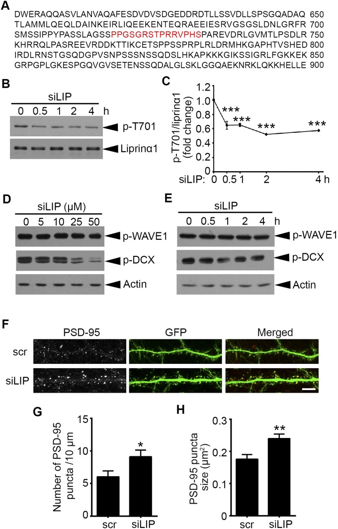
Fig. S4.
Liprinα1 siLIP promotes synaptic localization of PSD-95. (A) The amino acid sequence of liprinα1 from 601 to 900, with the sequence of the siLIP highlighted in red. (B and C) Liprinα1 siLIP inhibited liprinα1 phosphorylation after treatment for 30 min to 4 h. (B) Western blot analysis of phosphorylated (p-T701) and total liprinα1. Cultured neurons were incubated with siLIP (10 μM) for the indicated period at 14 DIV. (C) Quantification of liprinα1 phosphorylation, normalized to liprinα1 protein level. ***P < 0.001, one-way ANOVA with the Student–Newman–Keuls test; n = 3 independent experiments. (D and E) Liprinα1 siLIP (≤10 μM) did not alter the levels of p-WAVE1 (Ser310) or p-DCX (Ser297). (D) Cultured neurons were incubated with siLIP for 2 h at the indicated dosages. (E) Cultured neurons were incubated with siLIP (10 μM) for the indicated periods. (F–H) Liprinα1 siLIP treatment increased the density and size of PSD-95 puncta at synaptic regions. Cultured hippocampal neurons were transfected with GFP at 12 DIV and treated with siLIP (10 μM, 30 min) at 19 DIV. (F) Representative images of PSD-95 localization along dendrites treated with scr or siLIP. (Scale bar: 10 μm.) (G and H) Quantification analysis of PSD-95 puncta density and size after treatment with scr (G) and siLIP (H). *P < 0.05; **P < 0.01, Student’s t test; n = 15 and 12 dendrites for scr and siLIP, respectively. All data are mean ± SEM.

