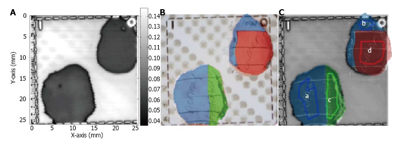
Figure 5.

An example terahertz image of excised cancerous, dysplastic and healthy colonic tissues. A: Example terahertz (THz) image of tissue containing healthy regions, dysplasia and cancerous tissue; B: The histology results (drawn onto a photographic image of the tissue samples); C: The histology results are overlaid on the THz image. In this example, regions a and b are normal tissue, c is dysplastic tissue and d is cancerous tissue[38] (printed with permission).
