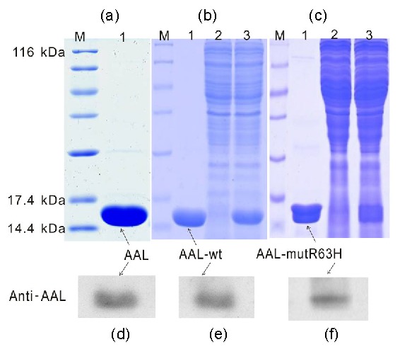
Fig. 1.

Purified AAL (a), AAL-wt (b), and AAL-mutR63H (c) applied to 15% SDS-PAGE and stained with Coomassie brilliant blue, and Western blot analysis of the purified AAL (d), AAL-wt (e), and AAL-mutR63H (f) using rabbit anti-AAL antibody
M, sizes (kD) of molecular weight markers; Lane 1, purified AAL, AAL-wt, or AAL-mutR63H; Land 2, flow-through; Land 3, crude extract AAL
