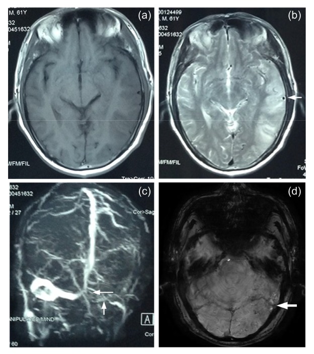
Fig. 1.

Magnetic resonance imaging (MRI), magnetic resonance venography (MRV), and susceptibility-weighted imaging (SWI)
(a) T1-weighted MRI; (b) T2-weighted MRI, and the arrow shows a flow void shadow of enlarged meningeal vessels in the left temporal lobe; (c) MRV, and the white arrows show a filling defect in the torcular and left transverse and sigmoid sinus; (d) SWI, and the white arrow shows hyperintensity in the left temporal lobe
