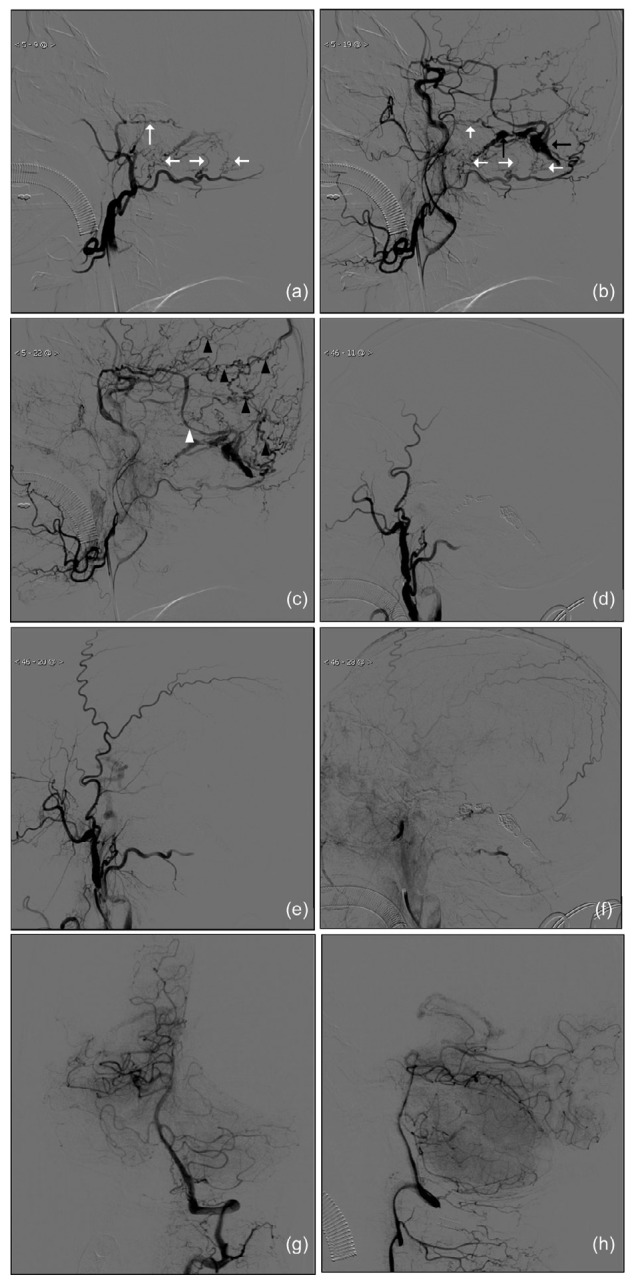
Fig. 2.

Preoperative and postoperative lateral digital subtraction angiography (DSA) images of the left external carotid artery (ECA) injection and preoperative DSA images of the left vertebral injection
Early (a), middle (b), and late (c) stages of the preoperative lateral DSA images; Early (d), middle (e), and late (f) stages of the postoperative lateral DSA images; Towne (g) and lateral (h) views of the preoperative DSA images of left vertebral injection. Black arrows show the early appearance of the left transverse and sigmoid sinus, confirming the diagnosis of a DAVF. White arrows show abnormally enlarged branches of the middle meningeal artery and occipital artery serving as feeders. The white arrowhead shows a thick vein at the left transverse sinus draining into the sphenoparietal sinus. Black arrowheads showed multiple retrograde cortical venous draining into the superior sagittal sinus
