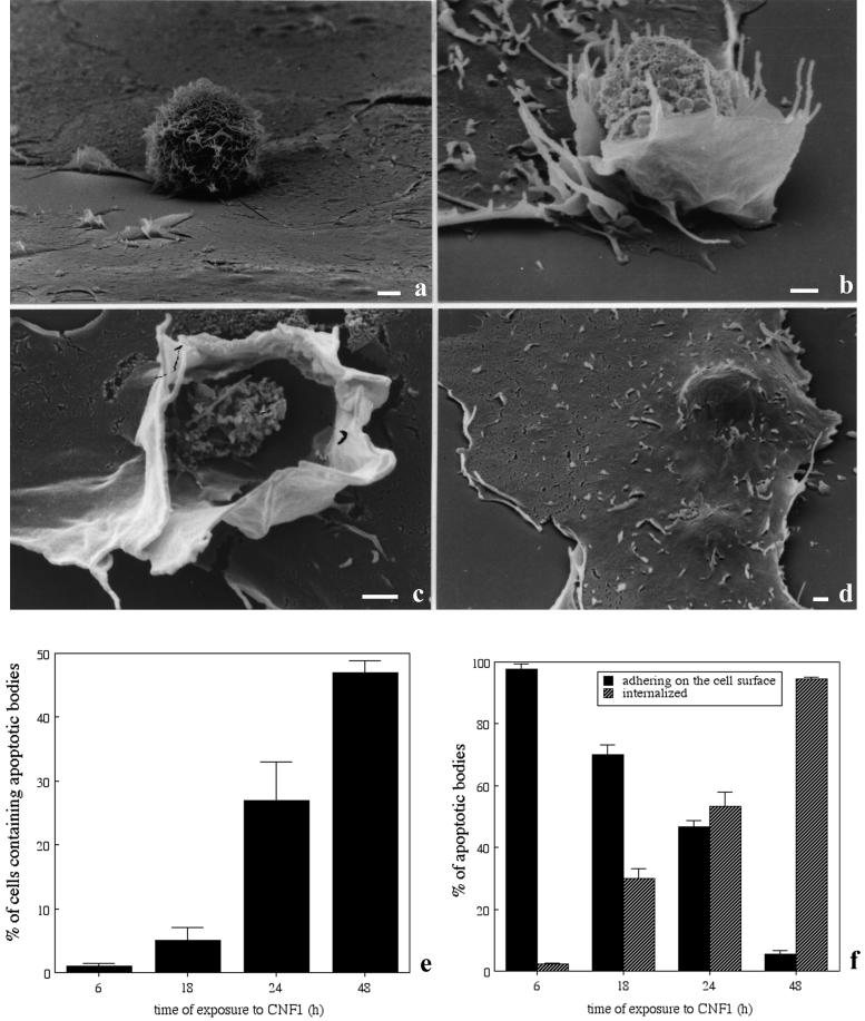
Figure 1.
Macropinocytosis of apoptotic bodies induced by CNF1 in epithelial cells. Scanning electron microscopy analysis of unstimulated epithelial cells (a) and cells exposed to 10−10 M CNF1 for 48 h (b–d). After overnight interaction with epithelial cells apoptotic bodies are contacted by filopodia and small ruffles (b), surrounded and enveloped (c) by larger ruffles, and internalized into the cytoplasm (d). (e) Percentage of cells containing apoptotic bodies increases by time of treatment with 10−10 M CNF1. (f) Percentage of apoptotic cells present on the epithelial cell surface decreases over time, whereas the number of internalized apoptotic bodies concomitantly increases. The results, reported as percentages (±SDs), are from four different experiments in each of which at least 500 cells (randomly chosen) were counted. Bar in a–d, 1 μm.

