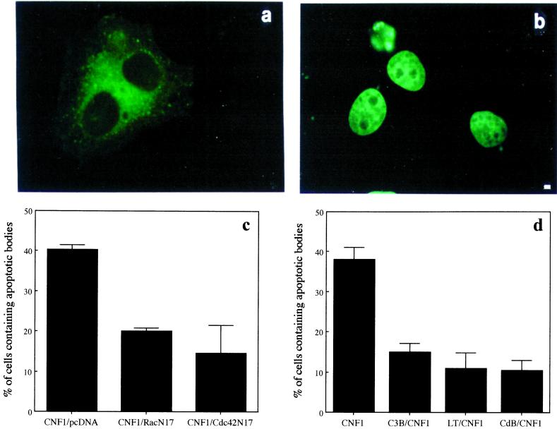
Figure 5.
Rho GTPases drive macropinocytosis in epithelial cells. (a and b) As an example of a transfected cell with an apoptotic body inside we show the fluorescence micrograph of an epithelial cell exposed to 10−10 M CNF1 for 24 h and then transfected with 1 μg of RacN17 encoding plasmid. Apoptotic bodies were added for 3 h before fixation. Hoechst staining (b) clearly evidences the presence of an apoptotic body in a transfected cell (a). (c) Graph showing the percentage of CNF1-treated epithelial cells able to macropinocytose apoptotic bodies once transfected with dominant negative forms of the Rho GTPases. (d) Graph showing the inhibition of CNF1-induced macropinocytotic activity by exposure to the Rho-inhibiting bacterial toxins C3B (1 μg/ml), LT (1 μg/ml), and CdB (0.5 μg/ml) for 3 h. The results reported as percentages (±SDs) of epithelial cells containing apoptotic bodies are from four different experiments in each of which at least 100 cells were counted. Bar in a and b, 1 μm.

