Figure 6.
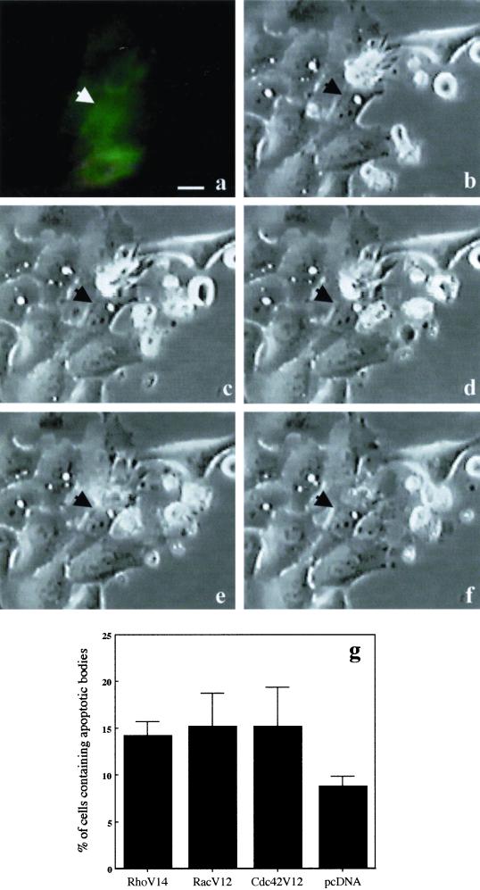
(a–f) Macropinocytosis of apoptotic bodies in HEp-2 cells transfected with RhoV14-GFP. Cells were transfected with 2.5 μg of RhoV14-GFP-encoding plasmid per Petri dish and then challenged with 5 × 10⁴ apoptotic cells. The interaction between a HEp-2 transfected cell (a) and an apoptotic cell was monitored by microcinematography. A selected field showing the dynamics of the capture and internalization processes was chosen (b–f). The selected micrographs were taken 10 min (b), 20 min (c), 1 h (d), 2 h (e), and 3 h (f) after the addition of apoptotic cells to the culture medium. Arrowheads indicate a transfected epithelial cell, which contacts and progressively internalizes an apoptotic cell. (g) Graph showing the percentage of control HEp-2 cells able to macropinocytose apoptotic bodies once transfected with 1 μg of dominant positive forms of Rho GTPase (RhoV14-RacV12-Cdc42V12 myc-tagged). In these cells a slight but significant increase in the macropinocytotic activity is evident with respect to cells transfected with the control plasmid (pcDNA). The results, reported as percentages (±SDs) of transfected epithelial cells containing apoptotic bodies, are from four different experiments in each of which at least 100 transfected cells were counted. Bar, 10 μm.

