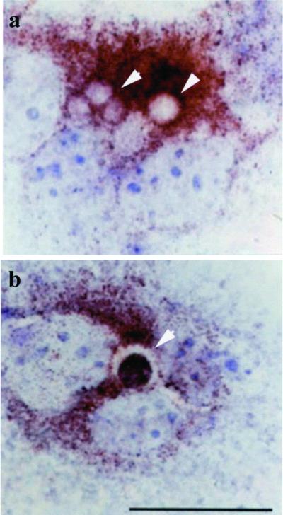
Figure 8.
Intracellular distribution of Lamp-1 in CNF1-treated epithelial cells. Immunocytochemistry analysis of epithelial cells exposed to 10−10 M CNF1 for 48 h (a and b). Arrowheads in a and b indicate vesicles stained with Lamp-1. In b is well evident an apoptotic body (stained with the terminal deoxynucleotidyl transferase dUTP nick-end labeling reaction) inside a Lamp-1-positive vesicle. The results obtained with Lamp-1 are from four separate experiments in each of which 100 cells were counted. Approximately 10% of Lamp-1-positive vesicles was found to contain apoptotic bodies. Bar, 10 μm.

